

For the pain physician, Urine Drug Testing (UDT) is not a tool for "catching" patients; it is a...
Comparative Review of H-Wave, F-Wave, and Somatic Evoked Potentials: Testing Setup, Anatomy, and Clinical Implications
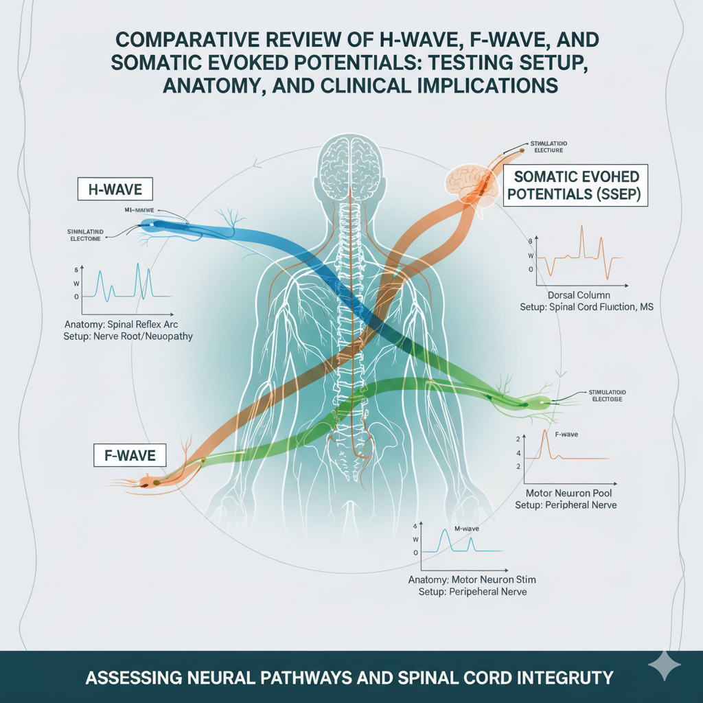
Neurophysiological testing plays a pivotal role in evaluating peripheral nerve function and central nervous system integrity. This review provides a comprehensive comparison of three common neurophysiological techniques: H-wave, F-wave, and somatic evoked potentials. Through elucidating their testing setups, anatomical correlates, clinical applications, and relevant clinical pearls, this article aims to facilitate a nuanced understanding of these diagnostic modalities among healthcare professionals.
Introduction: Neurophysiological testing encompasses a diverse array of techniques, each offering unique insights into peripheral nerve and central nervous system function. Understanding the differences between H-wave, F-wave, and somatic evoked potentials is essential for accurate diagnosis, localization of pathology, and monitoring treatment response.
H-Wave:
- Testing Setup: H-wave testing involves stimulating a peripheral nerve and recording the resulting Hoffman reflex (H-reflex) from a muscle innervated by the same nerve. The stimulus is typically delivered at a point along the nerve where the motor axons are absent, allowing selective activation of sensory fibers and subsequent reflex responses.
- Anatomy: The H-reflex reflects the excitability of the monosynaptic stretch reflex arc, involving the sensory nerve, spinal cord segment, and motor neuron pool. Its amplitude and latency provide indirect measures of peripheral nerve conduction velocity and spinal cord excitability.
- Clinical Applications: H-wave studies are valuable for assessing peripheral nerve function, spinal cord integrity, and the effects of neurological disorders or interventions on reflex pathways. They are particularly useful in evaluating conditions such as radiculopathy, peripheral neuropathy, and spinal cord injury.
- Clinical Pearls: Variations in H-wave parameters may occur due to factors such as age, gender, limb position, and level of muscle activation. Interpretation should consider these variables and compare findings to established normative data.
F-Wave:
- Testing Setup: F-wave testing involves stimulating a peripheral nerve and recording the resulting muscle action potentials (F-waves) evoked by antidromic activation of motor axons. F-waves represent the re-emergence of action potentials following antidromic conduction and subsequent orthodromic reactivation of motor neurons.
- Anatomy: F-waves provide insights into the excitability and conduction properties of motor nerve fibers, reflecting the integrity of proximal nerve segments, nerve roots, and spinal cord motor pathways.
- Clinical Applications: F-wave studies are valuable for evaluating nerve conduction velocity, detecting conduction block or temporal dispersion, localizing nerve lesions, and monitoring disease progression or treatment response in conditions such as neuropathy, radiculopathy, and motor neuron disorders.
- Clinical Pearls: F-wave parameters, including latency, amplitude, and persistence, may be influenced by factors such as limb temperature, electrode placement, and stimulus intensity. Standardized methodology and comparison with age-matched controls are essential for accurate interpretation.
Somatic Evoked Potentials:
- Testing Setup: Somatic evoked potentials involve delivering sensory or motor stimuli and recording the resulting electrical responses from the central nervous system, typically using scalp electrodes. Common modalities include sensory evoked potentials (SEPs), motor evoked potentials (MEPs), and auditory evoked potentials (AEPs).
- Anatomy: Somatic evoked potentials reflect the integrity and functional connectivity of sensory and motor pathways within the central nervous system, including the spinal cord, brainstem, and cerebral cortex.
- Clinical Applications: Somatic evoked potentials are used to assess sensory and motor function, localize lesions within the central nervous system, diagnose demyelinating disorders, monitor intraoperative neurophysiological integrity, and evaluate cognitive function in conditions such as multiple sclerosis, stroke, and spinal cord injury.
- Clinical Pearls: Signal acquisition, filtering, and artifact rejection are crucial for obtaining reliable somatic evoked potential recordings. Interpretation should consider age-related changes, neurological comorbidities, and concurrent medications affecting central nervous system function.
Conclusion: H-wave, F-wave, and somatic evoked potentials represent invaluable tools for evaluating peripheral nerve and central nervous system function across a spectrum of neurological conditions. Understanding their testing setups, anatomical correlates, clinical applications, and interpretation nuances is essential for healthcare professionals involved in neurophysiological testing and patient care. By integrating these modalities into clinical practice and leveraging their complementary insights, clinicians can enhance diagnostic accuracy, optimize treatment strategies, and improve outcomes for patients with neurological disorders.

Electrodiagnostic Medicine: A Review of NCS and EMG
Electrodiagnostic (EDX) medicine is a critical diagnostic tool in the fields of pain medicine, neurology, and physiatry. It is an extension of the physical examination used to evaluate the function of the peripheral nervous system, including motor and sensory nerves,...

Exploring Electromyography Findings: Insights into Polyphasic Waves, Positive Sharp Waves, and Fibrillations
Electromyography (EMG) serves as a vital diagnostic tool in assessing neuromuscular disorders by detecting electrical activity within muscles. This review delves into the main EMG findings, including polyphasic waves, positive sharp waves, fibrillations, and other...
Explore More
Toxicology and Urine Drug Testing (UDT)
For the pain physician, Urine Drug Testing (UDT) is not a tool for "catching" patients; it is a...
Adjuvant Medications—Alpha-2 Agonists and Steroids
In the pharmacological management of pain, "adjuvant" medications are those primarily indicated...
Topical Analgesics—Lidocaine, Capsaicin, and Compounded Creams
In the modern landscape of pain management, topical analgesics have transitioned from "ancillary"...